


 Foundational characteristics of cancer include proliferation, angiogenesis, migration, evasion of apoptosis, and cellular immortality. Find key markers for these cellular processes and antibodies to detect them.
 The SUMOplot™ Analysis Program predicts and scores sumoylation sites in your protein. SUMOylation is a post-translational modification involved in various cellular processes, such as nuclear-cytosolic transport, transcriptional regulation, apoptosis, protein stability, response to stress, and progression through the cell cycle.
 The Autophagy Receptor Motif Plotter predicts and scores autophagy receptor binding sites in your protein. Identifying proteins connected to this pathway is critical to understanding the role of autophagy in physiological as well as pathological processes such as development, differentiation, neurodegenerative diseases, stress, infection, and cancer.
Phospho-PAK1(T423) Antibody
Affinity Purified Rabbit Polyclonal Antibody (Pab)
- SPECIFICATION
- CITATIONS: 2
- PROTOCOLS
- BACKGROUND

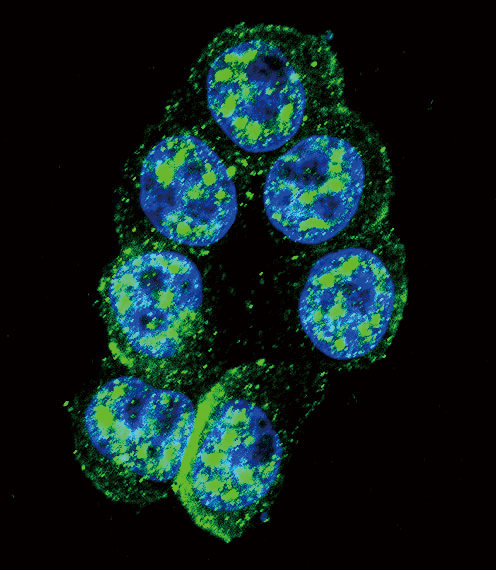
Application
| WB, IF, E |
|---|---|
| Primary Accession | Q13153 |
| Other Accession | Q8AXB4, Q62829, Q61036, O75914, Q64303, Q29502, Q8CIN4, Q13177, P35465, O88643, Q08E52 |
| Reactivity | Human |
| Predicted | Bovine, Mouse, Rat, Rabbit, Xenopus |
| Host | Rabbit |
| Clonality | Polyclonal |
| Isotype | Rabbit IgG |
| Calculated MW | 60647 Da |
| Gene ID | 5058 |
|---|---|
| Other Names | Serine/threonine-protein kinase PAK 1, Alpha-PAK, p21-activated kinase 1, PAK-1, p65-PAK, PAK1 |
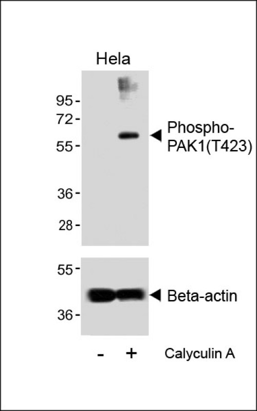
| Target/Specificity | This PAK1 Antibody is generated from rabbits immunized with a KLH conjugated synthetic phosphopeptide corresponding to amino acid residues surrounding T423 of human PAK1. |
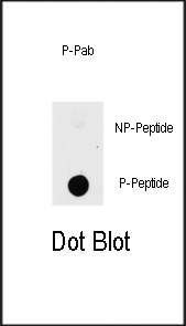
| Dilution | WB~~1:500 IF~~1:10~50 DB~~1:500 |
| Format | Purified polyclonal antibody supplied in PBS with 0.09% (W/V) sodium azide. This antibody is purified through a protein A column, followed by peptide affinity purification. |
| Storage | Maintain refrigerated at 2-8°C for up to 2 weeks. For long term storage store at -20°C in small aliquots to prevent freeze-thaw cycles. |
| Precautions | Phospho-PAK1(T423) Antibody is for research use only and not for use in diagnostic or therapeutic procedures. |
| Name | PAK1 {ECO:0000303|PubMed:8805275, ECO:0000312|HGNC:HGNC:8590} |
|---|---|
| Function | Protein kinase involved in intracellular signaling pathways downstream of integrins and receptor-type kinases that plays an important role in cytoskeleton dynamics, in cell adhesion, migration, proliferation, apoptosis, mitosis, and in vesicle-mediated transport processes (PubMed:10551809, PubMed:11896197, PubMed:12876277, PubMed:14585966, PubMed:15611088, PubMed:17726028, PubMed:17989089, PubMed:30290153). Can directly phosphorylate BAD and protects cells against apoptosis (By similarity). Activated by interaction with CDC42 and RAC1 (PubMed:8805275, PubMed:9528787). Functions as a GTPase effector that links the Rho-related GTPases CDC42 and RAC1 to the JNK MAP kinase pathway (PubMed:8805275, PubMed:9528787). Phosphorylates and activates MAP2K1, and thereby mediates activation of downstream MAP kinases (By similarity). Involved in the reorganization of the actin cytoskeleton, actin stress fibers and of focal adhesion complexes (PubMed:9395435, PubMed:9032240). Phosphorylates the tubulin chaperone TBCB and thereby plays a role in the regulation of microtubule biogenesis and organization of the tubulin cytoskeleton (PubMed:15831477). Plays a role in the regulation of insulin secretion in response to elevated glucose levels (PubMed:22669945). Part of a ternary complex that contains PAK1, DVL1 and MUSK that is important for MUSK-dependent regulation of AChR clustering during the formation of the neuromuscular junction (NMJ) (By similarity). Activity is inhibited in cells undergoing apoptosis, potentially due to binding of CDC2L1 and CDC2L2 (PubMed:12624090). Phosphorylates MYL9/MLC2 (By similarity). Phosphorylates RAF1 at 'Ser-338' and 'Ser-339' resulting in: activation of RAF1, stimulation of RAF1 translocation to mitochondria, phosphorylation of BAD by RAF1, and RAF1 binding to BCL2 (PubMed:11733498). Phosphorylates SNAI1 at 'Ser-246' promoting its transcriptional repressor activity by increasing its accumulation in the nucleus (PubMed:15833848). In podocytes, promotes NR3C2 nuclear localization (By similarity). Required for atypical chemokine receptor ACKR2-induced phosphorylation of LIMK1 and cofilin (CFL1) and for the up-regulation of ACKR2 from endosomal compartment to cell membrane, increasing its efficiency in chemokine uptake and degradation (PubMed:23633677). In synapses, seems to mediate the regulation of F- actin cluster formation performed by SHANK3, maybe through CFL1 phosphorylation and inactivation (By similarity). Plays a role in RUFY3-mediated facilitating gastric cancer cells migration and invasion (PubMed:25766321). In response to DNA damage, phosphorylates MORC2 which activates its ATPase activity and facilitates chromatin remodeling (PubMed:23260667). In neurons, plays a crucial role in regulating GABA(A) receptor synaptic stability and hence GABAergic inhibitory synaptic transmission through its role in F-actin stabilization (By similarity). In hippocampal neurons, necessary for the formation of dendritic spines and excitatory synapses; this function is dependent on kinase activity and may be exerted by the regulation of actomyosin contractility through the phosphorylation of myosin II regulatory light chain (MLC) (By similarity). Along with GIT1, positively regulates microtubule nucleation during interphase (PubMed:27012601). Phosphorylates FXR1, promoting its localization to stress granules and activity (PubMed:20417602). |
| Cellular Location | Cytoplasm. Cell junction, focal adhesion. Cell projection, lamellipodium. Cell membrane. Cell projection, ruffle membrane. Cell projection, invadopodium. Nucleus, nucleoplasm. Chromosome. Cytoplasm, cytoskeleton, microtubule organizing center, centrosome Note=Colocalizes with RUFY3, F-actin and other core migration components in invadopodia at the cell periphery (PubMed:25766321) Recruited to the cell membrane by interaction with CDC42 and RAC1 Recruited to focal adhesions upon activation. Colocalized with CIB1 within membrane ruffles during cell spreading upon readhesion to fibronectin. Upon DNA damage, translocates to the nucleoplasm when phosphorylated at Thr-212 where is co-recruited with MORC2 on damaged chromatin (PubMed:23260667). Localization to the centrosome does not depend upon the presence of gamma-tubulin (PubMed:27012601) Localization of the active, but not inactive, protein to the adhesions and edge of lamellipodia is mediated by interaction with GIT1 (PubMed:11896197). {ECO:0000250|UniProtKB:P35465, ECO:0000269|PubMed:11896197, ECO:0000269|PubMed:23260667, ECO:0000269|PubMed:25766321, ECO:0000269|PubMed:27012601} |
| Tissue Location | Overexpressed in gastric cancer cells and tissues (at protein level) (PubMed:25766321). |

Provided below are standard protocols that you may find useful for product applications.
Background
PAK proteins are critical effectors that link RhoGTPases to cytoskeleton reorganization and nuclear signaling. PAK proteins, a family of serine/threonine p21-activating kinases, include PAK1, PAK2, PAK3 and PAK4. These proteins serve as targets for the small GTP binding proteins Cdc42 and Rac and have been implicated in a wide range of biological activities. PAK1 regulates cell motility and morphology.
References
Li, W., et al., J. Biol. Chem. 279(31):32824-32831 (2004).
DerMardirossian, C., et al., Mol. Cell 15(1):117-127 (2004).
Weber, D.S., et al., Circ. Res. 94(9):1219-1226 (2004).
Balasenthil, S., et al., J. Biol. Chem. 279(2):1422-1428 (2004).
Wu, R.F., et al., J. Virol. 78(2):779-789 (2004).
If you have used an Abcepta product and would like to share how it has performed, please click on the "Submit Review" button and provide the requested information. Our staff will examine and post your review and contact you if needed.
If you have any additional inquiries please email technical services at tech@abcepta.com.
Ordering Information
Other Products
Shipping Information
