


 Foundational characteristics of cancer include proliferation, angiogenesis, migration, evasion of apoptosis, and cellular immortality. Find key markers for these cellular processes and antibodies to detect them.
 The SUMOplot™ Analysis Program predicts and scores sumoylation sites in your protein. SUMOylation is a post-translational modification involved in various cellular processes, such as nuclear-cytosolic transport, transcriptional regulation, apoptosis, protein stability, response to stress, and progression through the cell cycle.
 The Autophagy Receptor Motif Plotter predicts and scores autophagy receptor binding sites in your protein. Identifying proteins connected to this pathway is critical to understanding the role of autophagy in physiological as well as pathological processes such as development, differentiation, neurodegenerative diseases, stress, infection, and cancer.
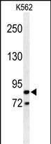
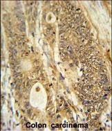
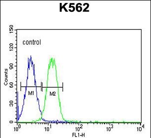
AVIL Antibody (N-term)
Affinity Purified Rabbit Polyclonal Antibody (Pab)
- SPECIFICATION
- CITATIONS
- PROTOCOLS
- BACKGROUND

Application
| FC, IHC-P, WB, E |
|---|---|
| Primary Accession | O75366 |
| Other Accession | O88398, Q9WU06 |
| Reactivity | Human |
| Predicted | Mouse, Rat |
| Host | Rabbit |
| Clonality | Polyclonal |
| Isotype | Rabbit IgG |
| Calculated MW | 92027 Da |
| Antigen Region | 176-204 aa |
| Gene ID | 10677 |
|---|---|
| Other Names | Advillin, p92, AVIL |
| Target/Specificity | This AVIL antibody is generated from rabbits immunized with a KLH conjugated synthetic peptide between 176-204 amino acids from the N-terminal region of human AVIL. |
| Dilution | WB~~1:1000 IHC-P~~1:50~100 FC~~1:10~50 |
| Format | Purified polyclonal antibody supplied in PBS with 0.09% (W/V) sodium azide. This antibody is purified through a protein A column, followed by peptide affinity purification. |
| Storage | Maintain refrigerated at 2-8°C for up to 2 weeks. For long term storage store at -20°C in small aliquots to prevent freeze-thaw cycles. |
| Precautions | AVIL Antibody (N-term) is for research use only and not for use in diagnostic or therapeutic procedures. |
| Name | AVIL (HGNC:14188) |
|---|---|
| Function | Ca(2+)-regulated actin-binding protein which plays an important role in actin bundling (PubMed:29058690). May have a unique function in the morphogenesis of neuronal cells which form ganglia. Required for SREC1-mediated regulation of neurite-like outgrowth. Plays a role in regenerative sensory axon outgrowth and remodeling processes after peripheral injury in neonates. Involved in the formation of long fine actin-containing filopodia-like structures in fibroblast. Plays a role in ciliogenesis. In podocytes, controls lamellipodia formation through the regulation of EGF-induced diacylglycerol generation by PLCE1 and ARP2/3 complex assembly (PubMed:29058690). |
| Cellular Location | Cytoplasm, cytoskeleton. Cell projection, lamellipodium. Cell junction, focal adhesion. Cell projection, neuron projection {ECO:0000250|UniProtKB:Q9WU06}. Cell projection, axon {ECO:0000250|UniProtKB:Q9WU06}. Note=In podocytes, present in the F- actin-enriched cell periphery that generates lamellipodia and focal adhesions. |
| Tissue Location | Most highly expressed in the small intestine and colonic lining. Weaker expression also detected in the thymus, prostate, testes and uterus (PubMed:12034507). Expressed in podocytes (at protein level) (PubMed:29058690). |

Thousands of laboratories across the world have published research that depended on the performance of antibodies from Abcepta to advance their research. Check out links to articles that cite our products in major peer-reviewed journals, organized by research category.
info@abcepta.com, and receive a free "I Love Antibodies" mug.

Provided below are standard protocols that you may find useful for product applications.
Background
AVIL is a member of the gelsolin/villin family of actin regulatory proteins. This protein has structural similarity to villin. It binds actin and may play a role in the development of neuronal cells that form ganglia.
References
Piana, S., et al. J. Mol. Biol. 375(2):460-470(2008)
Vermeulen, W., et al. Protein Sci. 13(5):1276-1287(2004)
Tumer, Z., et al. Gene 288 (1-2), 179-185 (2002)
If you have used an Abcepta product and would like to share how it has performed, please click on the "Submit Review" button and provide the requested information. Our staff will examine and post your review and contact you if needed.
If you have any additional inquiries please email technical services at tech@abcepta.com.

Ordering Information
Shipping Information
