


 Foundational characteristics of cancer include proliferation, angiogenesis, migration, evasion of apoptosis, and cellular immortality. Find key markers for these cellular processes and antibodies to detect them.
 The SUMOplot™ Analysis Program predicts and scores sumoylation sites in your protein. SUMOylation is a post-translational modification involved in various cellular processes, such as nuclear-cytosolic transport, transcriptional regulation, apoptosis, protein stability, response to stress, and progression through the cell cycle.
 The Autophagy Receptor Motif Plotter predicts and scores autophagy receptor binding sites in your protein. Identifying proteins connected to this pathway is critical to understanding the role of autophagy in physiological as well as pathological processes such as development, differentiation, neurodegenerative diseases, stress, infection, and cancer.
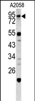
CALD1 Antibody (Center)
Purified Rabbit Polyclonal Antibody (Pab)

- SPECIFICATION
- CITATIONS
- PROTOCOLS
- BACKGROUND

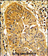
Application
| IHC-P, FC, WB, E |
|---|---|
| Primary Accession | Q05682 |
| Reactivity | Human |
| Host | Rabbit |
| Clonality | Polyclonal |
| Isotype | Rabbit IgG |
| Calculated MW | 93231 Da |
| Antigen Region | 428-457 aa |
| Gene ID | 800 |
|---|---|
| Other Names | Caldesmon, CDM, CALD1, CAD, CDM |
| Target/Specificity | This CALD1 antibody is generated from rabbits immunized with a KLH conjugated synthetic peptide between 428-457 amino acids from the Central region of human CALD1. |
| Dilution | WB~~1:1000 IHC-P~~1:10~50 FC~~1:10~50 |
| Format | Purified polyclonal antibody supplied in PBS with 0.09% (W/V) sodium azide. This antibody is prepared by Saturated Ammonium Sulfate (SAS) precipitation followed by dialysis against PBS. |
| Storage | Maintain refrigerated at 2-8°C for up to 2 weeks. For long term storage store at -20°C in small aliquots to prevent freeze-thaw cycles. |
| Precautions | CALD1 Antibody (Center) is for research use only and not for use in diagnostic or therapeutic procedures. |
| Name | CALD1 |
|---|---|
| Synonyms | CAD, CDM |
| Function | Actin- and myosin-binding protein implicated in the regulation of actomyosin interactions in smooth muscle and nonmuscle cells (could act as a bridge between myosin and actin filaments). Stimulates actin binding of tropomyosin which increases the stabilization of actin filament structure. In muscle tissues, inhibits the actomyosin ATPase by binding to F-actin. This inhibition is attenuated by calcium-calmodulin and is potentiated by tropomyosin. Interacts with actin, myosin, two molecules of tropomyosin and with calmodulin. Also plays an essential role during cellular mitosis and receptor capping. Involved in Schwann cell migration during peripheral nerve regeneration (By similarity). |
| Cellular Location | Cytoplasm, cytoskeleton {ECO:0000250|UniProtKB:P13505}. Cytoplasm, myofibril {ECO:0000250|UniProtKB:P13505}. Cytoplasm, cytoskeleton, stress fiber {ECO:0000250|UniProtKB:P13505}. Note=On thin filaments in smooth muscle and on stress fibers in fibroblasts (nonmuscle) {ECO:0000250|UniProtKB:P13505} |
| Tissue Location | High-molecular-weight caldesmon (isoform 1) is predominantly expressed in smooth muscles, whereas low-molecular-weight caldesmon (isoforms 2, 3, 4 and 5) are widely distributed in non-muscle tissues and cells. Not expressed in skeletal muscle or heart |

Thousands of laboratories across the world have published research that depended on the performance of antibodies from Abcepta to advance their research. Check out links to articles that cite our products in major peer-reviewed journals, organized by research category.
info@abcepta.com, and receive a free "I Love Antibodies" mug.

Provided below are standard protocols that you may find useful for product applications.
Background
CALD1 is a calmodulin- and actin-binding protein that plays an essential role in the regulation of smooth muscle and nonmuscle contraction. The conserved domain of this protein possesses the binding activities to Ca(2+)-calmodulin, actin, tropomyosin, myosin, and phospholipids. This protein is a potent inhibitor of the actin-tropomyosin activated myosin MgATPase, and serves as a mediating factor for Ca(2+)-dependent inhibition of smooth muscle contraction.
References
Yoshio,T., FEBS Lett. 581 (20), 3777-3782 (2007) Mani,R.S., Biochemistry 31 (47), 11896-11901 (1992)
If you have used an Abcepta product and would like to share how it has performed, please click on the "Submit Review" button and provide the requested information. Our staff will examine and post your review and contact you if needed.
If you have any additional inquiries please email technical services at tech@abcepta.com.

Ordering Information
Other Products
Shipping Information
