


 Foundational characteristics of cancer include proliferation, angiogenesis, migration, evasion of apoptosis, and cellular immortality. Find key markers for these cellular processes and antibodies to detect them.
 The SUMOplot™ Analysis Program predicts and scores sumoylation sites in your protein. SUMOylation is a post-translational modification involved in various cellular processes, such as nuclear-cytosolic transport, transcriptional regulation, apoptosis, protein stability, response to stress, and progression through the cell cycle.
 The Autophagy Receptor Motif Plotter predicts and scores autophagy receptor binding sites in your protein. Identifying proteins connected to this pathway is critical to understanding the role of autophagy in physiological as well as pathological processes such as development, differentiation, neurodegenerative diseases, stress, infection, and cancer.
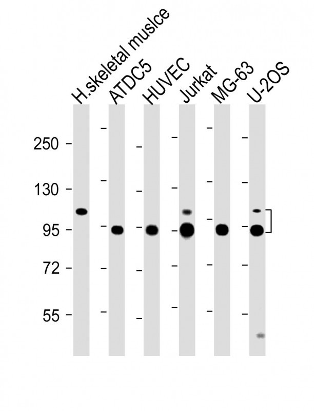
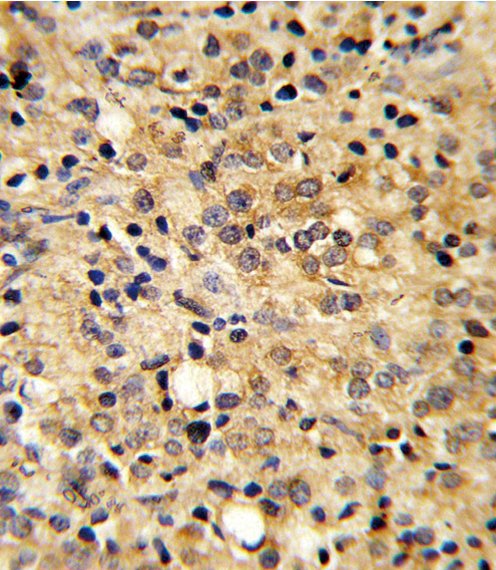
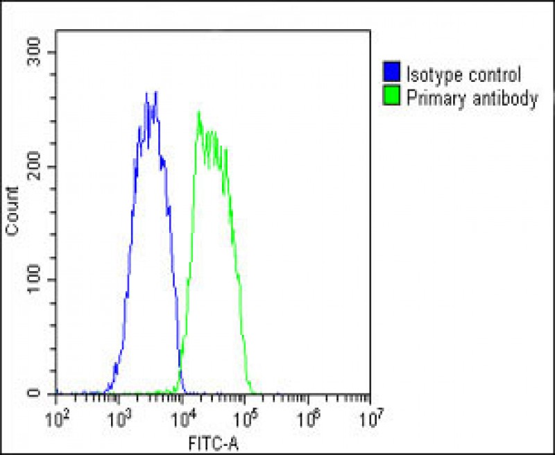
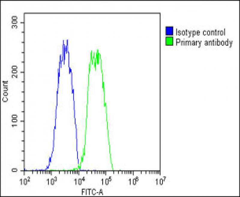
COMP Antibody (Center)
Affinity Purified Rabbit Polyclonal Antibody (Pab)

- SPECIFICATION
- CITATIONS
- PROTOCOLS
- BACKGROUND

Application
| IHC-P, WB, FC, E |
|---|---|
| Primary Accession | P49747 |
| Reactivity | Human |
| Host | Rabbit |
| Clonality | Polyclonal |
| Isotype | Rabbit IgG |
| Calculated MW | 82860 Da |
| Antigen Region | 314-343 aa |
| Gene ID | 1311 |
|---|---|
| Other Names | Cartilage oligomeric matrix protein, COMP, Thrombospondin-5, TSP5, COMP |
| Target/Specificity | This COMP antibody is generated from rabbits immunized with a KLH conjugated synthetic peptide between 314-343 amino acids from the Central region of human COMP. |
| Dilution | WB~~1:2000 IHC-P~~1:10~50 FC~~1:25 |
| Format | Purified polyclonal antibody supplied in PBS with 0.09% (W/V) sodium azide. This antibody is purified through a protein A column, followed by peptide affinity purification. |
| Storage | Maintain refrigerated at 2-8°C for up to 2 weeks. For long term storage store at -20°C in small aliquots to prevent freeze-thaw cycles. |
| Precautions | COMP Antibody (Center) is for research use only and not for use in diagnostic or therapeutic procedures. |
| Name | COMP (HGNC:2227) |
|---|---|
| Function | Plays a role in the structural integrity of cartilage via its interaction with other extracellular matrix proteins such as the collagens and fibronectin. Can mediate the interaction of chondrocytes with the cartilage extracellular matrix through interaction with cell surface integrin receptors (PubMed:16542502, PubMed:16051604). Could play a role in the pathogenesis of osteoarthritis (PubMed:16542502). Potent suppressor of apoptosis in both primary chondrocytes and transformed cells. Suppresses apoptosis by blocking the activation of caspase-3 and by inducing the IAP family of survival proteins (BIRC3, BIRC2, BIRC5 and XIAP) (PubMed:17993464). Essential for maintaining a vascular smooth muscle cells (VSMCs) contractile/differentiated phenotype under physiological and pathological stimuli. Maintains this phenotype of VSMCs by interacting with ITGA7 (By similarity). |
| Cellular Location | Secreted, extracellular space, extracellular matrix |
| Tissue Location | Abundantly expressed in the chondrocyte extracellular matrix, and is also found in bone, tendon, ligament and synovium and blood vessels. Increased amounts are produced during late stages of osteoarthritis in the area adjacent to the main defect |

Thousands of laboratories across the world have published research that depended on the performance of antibodies from Abcepta to advance their research. Check out links to articles that cite our products in major peer-reviewed journals, organized by research category.
info@abcepta.com, and receive a free "I Love Antibodies" mug.

Provided below are standard protocols that you may find useful for product applications.
Background
COMP is a noncollagenous extracellular matrix (ECM) protein. It consists of five identical glycoprotein subunits, each with EGF-like and calcium-binding (thrombospondin-like) domains. Oligomerization results from formation of a five-stranded coiled coil and disulfides. Binding to other ECM proteins such as collagen appears to depend on divalent cations.
References
Kim,H.J., et.al., Eur. J. Appl. Physiol. 105 (5), 765-770 (2009)
If you have used an Abcepta product and would like to share how it has performed, please click on the "Submit Review" button and provide the requested information. Our staff will examine and post your review and contact you if needed.
If you have any additional inquiries please email technical services at tech@abcepta.com.
Ordering Information
Other Products
Shipping Information
