


 Foundational characteristics of cancer include proliferation, angiogenesis, migration, evasion of apoptosis, and cellular immortality. Find key markers for these cellular processes and antibodies to detect them.
 The SUMOplot™ Analysis Program predicts and scores sumoylation sites in your protein. SUMOylation is a post-translational modification involved in various cellular processes, such as nuclear-cytosolic transport, transcriptional regulation, apoptosis, protein stability, response to stress, and progression through the cell cycle.
 The Autophagy Receptor Motif Plotter predicts and scores autophagy receptor binding sites in your protein. Identifying proteins connected to this pathway is critical to understanding the role of autophagy in physiological as well as pathological processes such as development, differentiation, neurodegenerative diseases, stress, infection, and cancer.
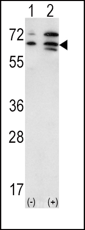
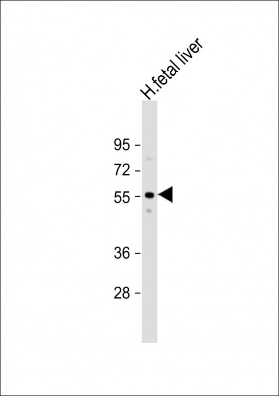
ACVR2A Antibody (N-term)
Purified Rabbit Polyclonal Antibody (Pab)

- SPECIFICATION
- CITATIONS
- PROTOCOLS
- BACKGROUND

Application
| WB, E |
|---|---|
| Primary Accession | P27037 |
| Other Accession | P38444, P27038, Q90669, Q28043, Q28560 |
| Reactivity | Human, Mouse |
| Predicted | Bovine, Chicken, Rat, Sheep |
| Host | Rabbit |
| Clonality | Polyclonal |
| Isotype | Rabbit IgG |
| Calculated MW | 57848 Da |
| Antigen Region | 2-29 aa |
| Gene ID | 92 |
|---|---|
| Other Names | Activin receptor type-2A, Activin receptor type IIA, ACTR-IIA, ACTRIIA, ACVR2A, ACVR2 |
| Target/Specificity | This ACVR2A antibody is generated from rabbits immunized with a KLH conjugated synthetic peptide between 2-29 amino acids from the N-terminal region of human ACVR2A. |
| Dilution | WB~~1:2000 |
| Format | Purified polyclonal antibody supplied in PBS with 0.09% (W/V) sodium azide. This antibody is prepared by Saturated Ammonium Sulfate (SAS) precipitation followed by dialysis against PBS. |
| Storage | Maintain refrigerated at 2-8°C for up to 2 weeks. For long term storage store at -20°C in small aliquots to prevent freeze-thaw cycles. |
| Precautions | ACVR2A Antibody (N-term) is for research use only and not for use in diagnostic or therapeutic procedures. |
| Name | ACVR2A (HGNC:173) |
|---|---|
| Synonyms | ACVR2 |
| Function | On ligand binding, forms a receptor complex consisting of two type II and two type I transmembrane serine/threonine kinases. Type II receptors phosphorylate and activate type I receptors which autophosphorylate, then bind and activate SMAD transcriptional regulators. Receptor for activin A, activin B and inhibin A (PubMed:17911401). Mediates induction of adipogenesis by GDF6 (By similarity). |
| Cellular Location | Cell membrane {ECO:0000250|UniProtKB:P27038}; Single-pass type I membrane protein |

Thousands of laboratories across the world have published research that depended on the performance of antibodies from Abcepta to advance their research. Check out links to articles that cite our products in major peer-reviewed journals, organized by research category.
info@abcepta.com, and receive a free "I Love Antibodies" mug.

Provided below are standard protocols that you may find useful for product applications.
Background
ACVR2A is an activin A type II receptor. Activins are dimeric growth and differentiation factors which belong to the transforming growth factor-beta (TGF-beta) superfamily of structurally related signaling proteins. Activins signal through a heteromeric complex of receptor serine kinases which include at least two type I (I and IB) and two type II (II and IIB) receptors. These receptors are all transmembrane proteins, composed of a ligand-binding extracellular domain with cysteine-rich region, a transmembrane domain, and a cytoplasmic domain with predicted serine/threonine specificity. Type I receptors are essential for signaling; and type II receptors are required for binding ligands and for expression of type I receptors. Type I and II receptors form a stable complex after ligand binding, resulting in phosphorylation of type I receptors by type II receptors. Type II receptors are considered to be constitutively active kinases.
References
Jung, B., et al., Gastroenterology 126(3):654-659 (2004).
Martins da Silva, S.J., et al., Dev. Biol. 266(2):334-345 (2004).
Olaru, A., et al., Lab. Invest. 83(12):1867-1871 (2003).
Casagrandi, D., et al., Mol. Hum. Reprod. 9(4):199-203 (2003).
Greenwald, J., et al., Mol. Cell 11(3):605-617 (2003).
If you have used an Abcepta product and would like to share how it has performed, please click on the "Submit Review" button and provide the requested information. Our staff will examine and post your review and contact you if needed.
If you have any additional inquiries please email technical services at tech@abcepta.com.
Ordering Information
Other Products
Shipping Information
