


 Foundational characteristics of cancer include proliferation, angiogenesis, migration, evasion of apoptosis, and cellular immortality. Find key markers for these cellular processes and antibodies to detect them.
 The SUMOplot™ Analysis Program predicts and scores sumoylation sites in your protein. SUMOylation is a post-translational modification involved in various cellular processes, such as nuclear-cytosolic transport, transcriptional regulation, apoptosis, protein stability, response to stress, and progression through the cell cycle.
 The Autophagy Receptor Motif Plotter predicts and scores autophagy receptor binding sites in your protein. Identifying proteins connected to this pathway is critical to understanding the role of autophagy in physiological as well as pathological processes such as development, differentiation, neurodegenerative diseases, stress, infection, and cancer.
> home > Products > Primary Antibodies > Antibody Collections > Anti-Kinome Antibodies > BLK Antibody (N-term)
BLK Antibody (N-term)
Purified Rabbit Polyclonal Antibody (Pab)

- SPECIFICATION
- CITATIONS
- PROTOCOLS
- BACKGROUND

Application
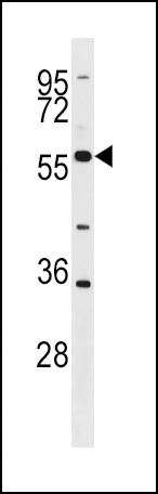
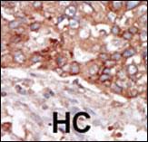
| IHC-P, WB, E |
|---|---|
| Primary Accession | P51451 |
| Reactivity | Human, Mouse |
| Host | Rabbit |
| Clonality | Polyclonal |
| Isotype | Rabbit IgG |
| Calculated MW | 57706 Da |
| Antigen Region | 1-30 aa |
| Gene ID | 640 |
|---|---|
| Other Names | Tyrosine-protein kinase Blk, B lymphocyte kinase, p55-Blk, BLK |
| Target/Specificity | This BLK antibody is generated from rabbits immunized with a KLH conjugated synthetic peptide between 1-30 amino acids from the N-terminal region of human BLK. |
| Dilution | WB~~1:1000 IHC-P~~1:50~100 |
| Format | Purified polyclonal antibody supplied in PBS with 0.09% (W/V) sodium azide. This antibody is purified through a protein A column, followed by peptide affinity purification. |
| Storage | Maintain refrigerated at 2-8°C for up to 2 weeks. For long term storage store at -20°C in small aliquots to prevent freeze-thaw cycles. |
| Precautions | BLK Antibody (N-term) is for research use only and not for use in diagnostic or therapeutic procedures. |
| Name | BLK |
|---|---|
| Function | Non-receptor tyrosine kinase involved in B-lymphocyte development, differentiation and signaling (By similarity). B-cell receptor (BCR) signaling requires a tight regulation of several protein tyrosine kinases and phosphatases, and associated coreceptors (By similarity). Binding of antigen to the B-cell antigen receptor (BCR) triggers signaling that ultimately leads to B-cell activation (By similarity). Signaling through BLK plays an important role in transmitting signals through surface immunoglobulins and supports the pro-B to pre-B transition, as well as the signaling for growth arrest and apoptosis downstream of B-cell receptor (By similarity). Specifically binds and phosphorylates CD79A at 'Tyr-188'and 'Tyr-199', as well as CD79B at 'Tyr-196' and 'Tyr-207' (By similarity). Also phosphorylates the immunoglobulin G receptors FCGR2A, FCGR2B and FCGR2C (PubMed:8756631). With FYN and LYN, plays an essential role in pre-B- cell receptor (pre-BCR)-mediated NF-kappa-B activation (By similarity). Also contributes to BTK activation by indirectly stimulating BTK intramolecular autophosphorylation (By similarity). In pancreatic islets, acts as a modulator of beta-cells function through the up- regulation of PDX1 and NKX6-1 and consequent stimulation of insulin secretion in response to glucose (PubMed:19667185). Phosphorylates CGAS, promoting retention of CGAS in the cytosol (PubMed:30356214). |
| Cellular Location | Cell membrane; Lipid-anchor. Note=Present and active in lipid rafts. Membrane location is required for the phosphorylation of CD79A and CD79B (By similarity). |
| Tissue Location | Expressed in lymphatic organs, pancreatic islets, Leydig cells, striate ducts of salivary glands and hair follicles |
Research Areas

Abcepta welcomes feedback from its customers.
If you have used an Abcepta product and would like to share how it has performed, please click on the "Submit Review" button and provide the requested information. Our staff will examine and post your review and contact you if needed.
If you have any additional inquiries please email technical services at tech@abcepta.com.
$ 192.50

Cat# AP7697a
Ordering Information
United States
AlbaniaAustraliaAustriaBelgiumBosnia & HerzegovinaBrazilBulgariaCanadaCentral AmericaChinaCroatiaCyprusCzech RepublicDenmarkEstoniaFinlandFranceGermanyGreeceHong KongHungaryIcelandIndiaIndonesiaIrelandIsraelItalyJapanLatviaLithuaniaLuxembourgMacedoniaMalaysiaMaltaNetherlandsNew ZealandNorwayPakistanPolandPortugalRomaniaSerbiaSingaporeSlovakiaSloveniaSouth AfricaSouth KoreaSpainSwedenSwitzerlandTaiwanTurkeyUnited KingdomUnited StatesVietnamWorldwideOthersMexico
USA Headquarters
(888) 735-7227 / (858) 622-0099 or (858) 875-1900
Other Products
Shipping Information
Domestic orders (in stock items)
Shipped out the same day. Orders placed after 1 PM (PST) will ship out the next business day.
International orders
Contact your local distributors
