


 Foundational characteristics of cancer include proliferation, angiogenesis, migration, evasion of apoptosis, and cellular immortality. Find key markers for these cellular processes and antibodies to detect them.
 The SUMOplot™ Analysis Program predicts and scores sumoylation sites in your protein. SUMOylation is a post-translational modification involved in various cellular processes, such as nuclear-cytosolic transport, transcriptional regulation, apoptosis, protein stability, response to stress, and progression through the cell cycle.
 The Autophagy Receptor Motif Plotter predicts and scores autophagy receptor binding sites in your protein. Identifying proteins connected to this pathway is critical to understanding the role of autophagy in physiological as well as pathological processes such as development, differentiation, neurodegenerative diseases, stress, infection, and cancer.
Activin A Receptor Type IB (ACVR1B) Antibody (N-term)
Purified Rabbit Polyclonal Antibody (Pab)
- SPECIFICATION
- CITATIONS
- PROTOCOLS
- BACKGROUND

Application
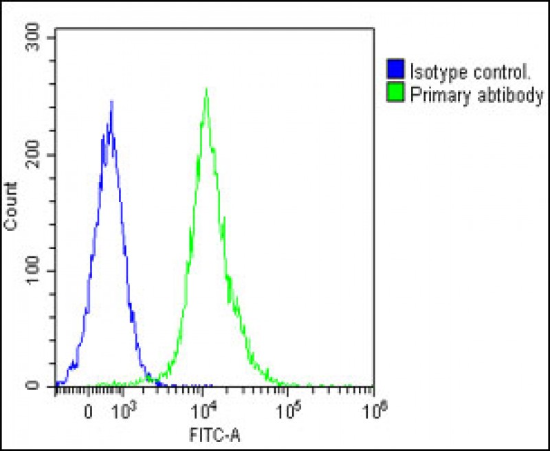
| WB, FC, E |
|---|---|
| Primary Accession | P36896 |
| Other Accession | P80202 |
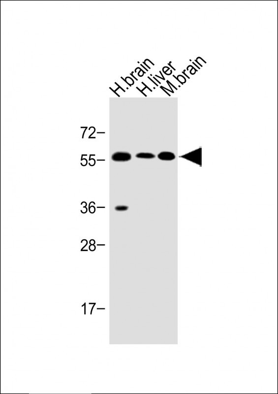
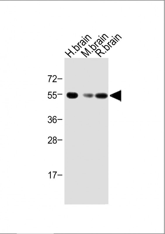
| Reactivity | Human, Mouse, Rat |
| Predicted | Rat |
| Host | Rabbit |
| Clonality | Polyclonal |
| Isotype | Rabbit IgG |
| Calculated MW | 56807 Da |
| Antigen Region | 39-68 aa |
| Gene ID | 91 |
|---|---|
| Other Names | Activin receptor type-1B, Activin receptor type IB, ACTR-IB, Activin receptor-like kinase 4, ALK-4, Serine/threonine-protein kinase receptor R2, SKR2, ACVR1B, ACVRLK4, ALK4 |
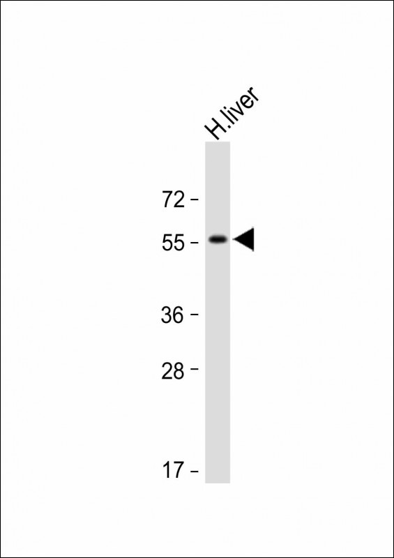
| Target/Specificity | This Activin A Receptor Type IB (ACVR1B) antibody is generated from rabbits immunized with a KLH conjugated synthetic peptide between 39-68 amino acids from the N-terminal region of human Activin A Receptor Type IB (ACVR1B). |
| Dilution | WB~~1:1000 FC~~1:25 |
| Format | Purified polyclonal antibody supplied in PBS with 0.09% (W/V) sodium azide. This antibody is purified through a protein A column, followed by peptide affinity purification. |
| Storage | Maintain refrigerated at 2-8°C for up to 2 weeks. For long term storage store at -20°C in small aliquots to prevent freeze-thaw cycles. |
| Precautions | Activin A Receptor Type IB (ACVR1B) Antibody (N-term) is for research use only and not for use in diagnostic or therapeutic procedures. |
| Name | ACVR1B |
|---|---|
| Synonyms | ACVRLK4, ALK4 |
| Function | Transmembrane serine/threonine kinase activin type-1 receptor forming an activin receptor complex with activin receptor type-2 (ACVR2A or ACVR2B). Transduces the activin signal from the cell surface to the cytoplasm and is thus regulating a many physiological and pathological processes including neuronal differentiation and neuronal survival, hair follicle development and cycling, FSH production by the pituitary gland, wound healing, extracellular matrix production, immunosuppression and carcinogenesis. Activin is also thought to have a paracrine or autocrine role in follicular development in the ovary. Within the receptor complex, type-2 receptors (ACVR2A and/or ACVR2B) act as a primary activin receptors whereas the type-1 receptors like ACVR1B act as downstream transducers of activin signals. Activin binds to type-2 receptor at the plasma membrane and activates its serine- threonine kinase. The activated receptor type-2 then phosphorylates and activates the type-1 receptor such as ACVR1B. Once activated, the type- 1 receptor binds and phosphorylates the SMAD proteins SMAD2 and SMAD3, on serine residues of the C-terminal tail. Soon after their association with the activin receptor and subsequent phosphorylation, SMAD2 and SMAD3 are released into the cytoplasm where they interact with the common partner SMAD4. This SMAD complex translocates into the nucleus where it mediates activin-induced transcription. Inhibitory SMAD7, which is recruited to ACVR1B through FKBP1A, can prevent the association of SMAD2 and SMAD3 with the activin receptor complex, thereby blocking the activin signal. Activin signal transduction is also antagonized by the binding to the receptor of inhibin-B via the IGSF1 inhibin coreceptor. ACVR1B also phosphorylates TDP2. |
| Cellular Location | Cell membrane; Single-pass type I membrane protein |
| Tissue Location | Expressed in many tissues, most strongly in kidney, pancreas, brain, lung, and liver |

Thousands of laboratories across the world have published research that depended on the performance of antibodies from Abcepta to advance their research. Check out links to articles that cite our products in major peer-reviewed journals, organized by research category.
info@abcepta.com, and receive a free "I Love Antibodies" mug.

Provided below are standard protocols that you may find useful for product applications.
Background
Activins are dimeric growth and differentiation factors which belong to the transforming growth factor-beta (TGF-beta) superfamily of structurally related signaling proteins. Activins signal through a heteromeric complex of receptor serine kinases which include at least two type I (I and IB) and two type II (II and IIB) receptors. These receptors are all transmembrane proteins, composed of a ligand-binding extracellular domain with a cysteine-rich region, a transmembrane domain, and a cytoplasmic domain with predicted serine/threonine specificity. Type I receptors are essential for signaling, and type II receptors are required for binding ligands and for expression of type I receptors. Type I and II receptors form a stable complex after ligand binding, resulting in phosphorylation of type I receptors by type II receptors. The gene for ACVR1B (activin A type IB receptor) is composed of 11 exons. Alternative splicing and alternative polyadenylation result in 3 fully described transcript variants. The mRNA expression of variants 1, 2, and 3 is confirmed, and a potential fourth variant contains an alternative exon 8 and lacks exons 9 through 11, but its mRNA expression has not been confirmed.
References
Harrison, C.A., et al., J. Biol. Chem. 278(23):21129-21135 (2003).
Mukasa, C., et al., Endocrinology 144(4):1603-1611 (2003).
Danila, D.C., et al., J. Clin. Endocrinol. Metab. 87(10):4741-4746 (2002).
Schneider-Kolsky, M.E., et al., Placenta 23(4):294-302 (2002).
Roijer, E., et al., Mamm. Genome 9(3):266-268 (1998).
If you have used an Abcepta product and would like to share how it has performed, please click on the "Submit Review" button and provide the requested information. Our staff will examine and post your review and contact you if needed.
If you have any additional inquiries please email technical services at tech@abcepta.com.

Ordering Information
Other Products
Shipping Information
