


 Foundational characteristics of cancer include proliferation, angiogenesis, migration, evasion of apoptosis, and cellular immortality. Find key markers for these cellular processes and antibodies to detect them.
 The SUMOplot™ Analysis Program predicts and scores sumoylation sites in your protein. SUMOylation is a post-translational modification involved in various cellular processes, such as nuclear-cytosolic transport, transcriptional regulation, apoptosis, protein stability, response to stress, and progression through the cell cycle.
 The Autophagy Receptor Motif Plotter predicts and scores autophagy receptor binding sites in your protein. Identifying proteins connected to this pathway is critical to understanding the role of autophagy in physiological as well as pathological processes such as development, differentiation, neurodegenerative diseases, stress, infection, and cancer.
PI3KR1 Antibody (N-term L11)
Purified Rabbit Polyclonal Antibody (Pab)

- SPECIFICATION
- CITATIONS: 1
- PROTOCOLS
- BACKGROUND

Application
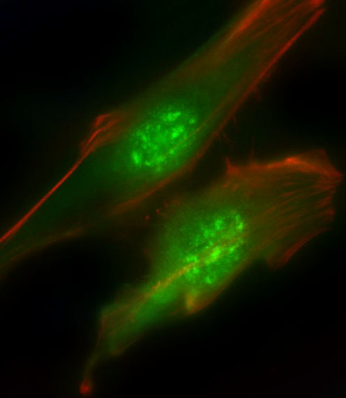
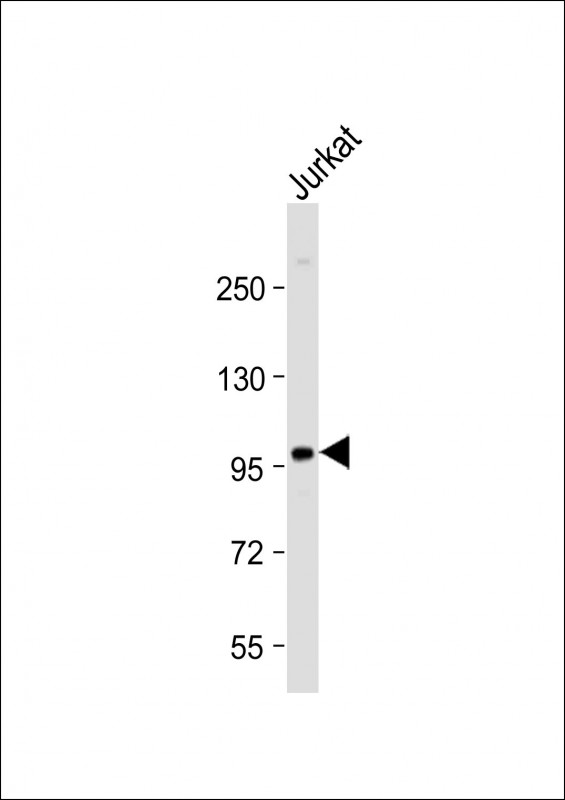
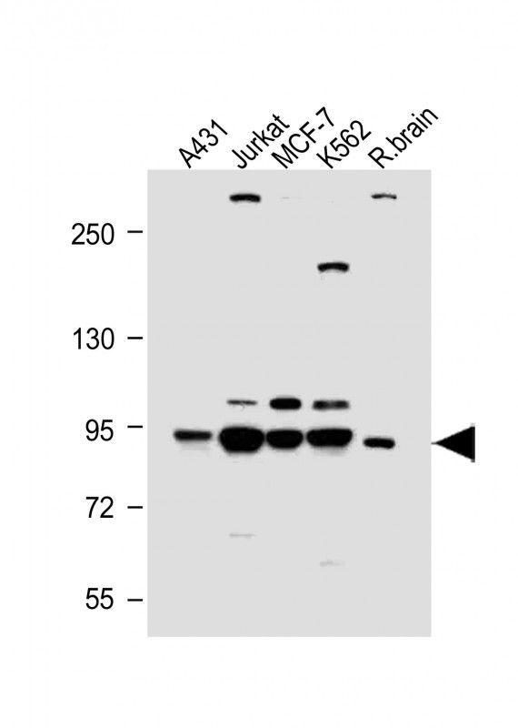
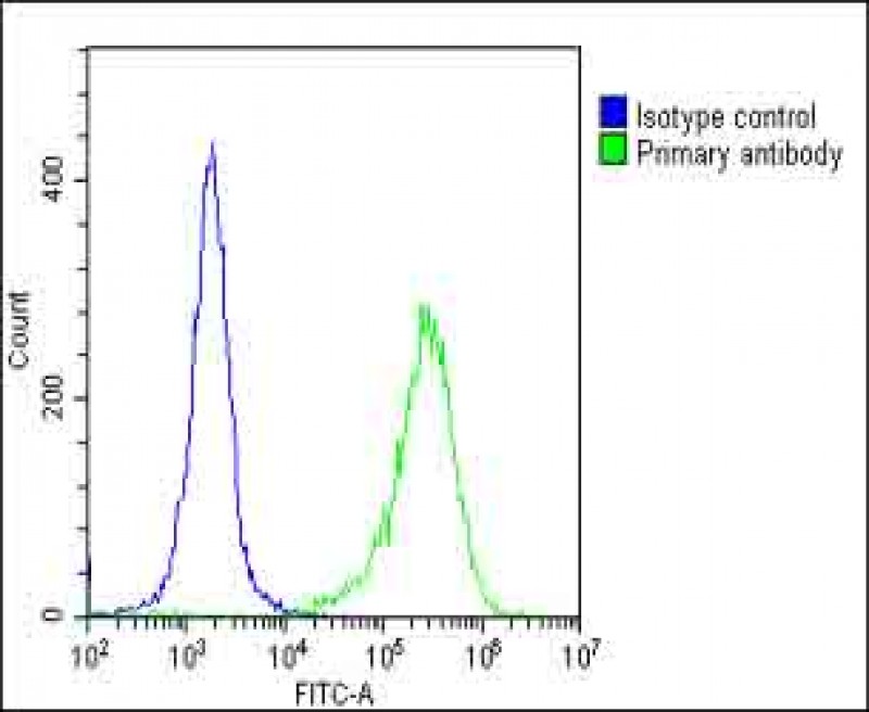
| IF, WB, FC, IHC-P-Leica, E |
|---|---|
| Primary Accession | P27986 |
| Other Accession | Q63787, P26450, P23727 |
| Reactivity | Human, Rat |
| Predicted | Bovine, Mouse |
| Host | Rabbit |
| Clonality | Polyclonal |
| Isotype | Rabbit IgG |
| Calculated MW | 83598 Da |
| Antigen Region | 1-30 aa |
| Gene ID | 5295 |
|---|---|
| Other Names | Phosphatidylinositol 3-kinase regulatory subunit alpha, PI3-kinase regulatory subunit alpha, PI3K regulatory subunit alpha, PtdIns-3-kinase regulatory subunit alpha, Phosphatidylinositol 3-kinase 85 kDa regulatory subunit alpha, PI3-kinase subunit p85-alpha, PtdIns-3-kinase regulatory subunit p85-alpha, PIK3R1, GRB1 |
| Target/Specificity | This PI3KR1 antibody is generated from rabbits immunized with a KLH conjugated synthetic peptide between 1-30 amino acids from the N-terminal region of human PI3KR1. |
| Dilution | IF~~1:25 WB~~1:2000 IHC-P-Leica~~1:500 FC~~1:25 |
| Format | Purified polyclonal antibody supplied in PBS with 0.09% (W/V) sodium azide. This antibody is purified through a protein A column, followed by peptide affinity purification. |
| Storage | Maintain refrigerated at 2-8°C for up to 2 weeks. For long term storage store at -20°C in small aliquots to prevent freeze-thaw cycles. |
| Precautions | PI3KR1 Antibody (N-term L11) is for research use only and not for use in diagnostic or therapeutic procedures. |
| Name | PIK3R1 |
|---|---|
| Synonyms | GRB1 |
| Function | Binds to activated (phosphorylated) protein-Tyr kinases, through its SH2 domain, and acts as an adapter, mediating the association of the p110 catalytic unit to the plasma membrane. Necessary for the insulin-stimulated increase in glucose uptake and glycogen synthesis in insulin-sensitive tissues. Plays an important role in signaling in response to FGFR1, FGFR2, FGFR3, FGFR4, KITLG/SCF, KIT, PDGFRA and PDGFRB. Likewise, plays a role in ITGB2 signaling (PubMed:17626883, PubMed:19805105, PubMed:7518429). Modulates the cellular response to ER stress by promoting nuclear translocation of XBP1 isoform 2 in a ER stress- and/or insulin-dependent manner during metabolic overloading in the liver and hence plays a role in glucose tolerance improvement (PubMed:20348923). |
| Tissue Location | Isoform 2 is expressed in skeletal muscle and brain, and at lower levels in kidney and cardiac muscle. Isoform 2 and isoform 4 are present in skeletal muscle (at protein level) |

Provided below are standard protocols that you may find useful for product applications.
Background
Phosphatidylinositol 3-kinase phosphorylates the inositol ring of phosphatidylinositol at the 3-prime position. The enzyme comprises a 110 kD catalytic subunit and a regulatory subunit of either 85, 55, or 50 kD. This gene encodes the 85 kD regulatory subunit. Phosphatidylinositol 3-kinase plays an important role in the metabolic actions of insulin, and a mutation in this gene has been associated with insulin resistance.
References
Kobayashi, H., et al., J. Biol. Chem. 279(8):6371-6379 (2004).
Liu, H., et al., J. Cell Biol. 164(4):603-612 (2004).
Sun, M., et al., J. Biol. Chem. 278(44):42992-43000 (2003).
Khan, N.A., et al., J. Neurovirol. 9(6):584-593 (2003).
Lee, H.Y., et al., J. Biol. Chem. 278(26):23630-23638 (2003).
If you have used an Abcepta product and would like to share how it has performed, please click on the "Submit Review" button and provide the requested information. Our staff will examine and post your review and contact you if needed.
If you have any additional inquiries please email technical services at tech@abcepta.com.
Ordering Information
Other Products
Shipping Information
