


 Foundational characteristics of cancer include proliferation, angiogenesis, migration, evasion of apoptosis, and cellular immortality. Find key markers for these cellular processes and antibodies to detect them.
 The SUMOplot™ Analysis Program predicts and scores sumoylation sites in your protein. SUMOylation is a post-translational modification involved in various cellular processes, such as nuclear-cytosolic transport, transcriptional regulation, apoptosis, protein stability, response to stress, and progression through the cell cycle.
 The Autophagy Receptor Motif Plotter predicts and scores autophagy receptor binding sites in your protein. Identifying proteins connected to this pathway is critical to understanding the role of autophagy in physiological as well as pathological processes such as development, differentiation, neurodegenerative diseases, stress, infection, and cancer.
AIF Antibody
- SPECIFICATION
- CITATIONS
- PROTOCOLS
- BACKGROUND

Application
| WB, IHC-P, E |
|---|---|
| Primary Accession | O95381 |
| Other Accession | O95381, 50400606 |
| Reactivity | Human, Mouse, Rat |
| Host | Rabbit |
| Clonality | Polyclonal |
| Isotype | IgG |
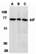
| Calculated MW | Predicted: 27, 36, 67 kDa Observed: 71 kDa |
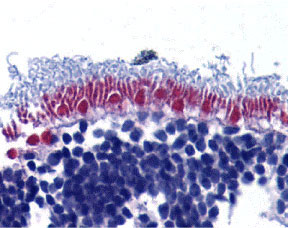
| Application Notes | AIF antibody can be used for detection of AIF by Western blot at 1 µg/mL. Antibody can also be used for immunohistochemistry starting at 10 µg/mL. |
| Gene ID | 10256 |
|---|---|
| Other Names | AIF Antibody: CNK, KSR, CNK1, Connector enhancer of kinase suppressor of ras 1, CNK homolog protein 1, Connector enhancer of KSR 1, connector enhancer of kinase suppressor of Ras 1 |
| Target/Specificity | CNKSR1; At least five isoforms of AIF are known to exist; this antibody will detect all isoforms except isoform 5. |
| Reconstitution & Storage | AIF antibody can be stored at 4℃ for three months and -20℃, stable for up to one year. As with all antibodies care should be taken to avoid repeated freeze thaw cycles. Antibodies should not be exposed to prolonged high temperatures. |
| Precautions | AIF Antibody is for research use only and not for use in diagnostic or therapeutic procedures. |

Thousands of laboratories across the world have published research that depended on the performance of antibodies from Abcepta to advance their research. Check out links to articles that cite our products in major peer-reviewed journals, organized by research category.
info@abcepta.com, and receive a free "I Love Antibodies" mug.

Provided below are standard protocols that you may find useful for product applications.
Background
AIF Antibody: Apoptosis is characterized by several morphological nuclear changes including chromatin condensation and nuclear fragmentation. These changes are triggered by the activation of members of caspase family, caspase activated DNase, and several novel proteins. A novel gene, the product of which causes chromatin condensation and DNA fragmentation, was recently identified, cloned, and designated apoptosis inducing factor (AIF). Like the critical molecules, cytochrome c and caspase-9, in apoptosis, AIF localizes in mitochondria. AIF translocates to the nucleus when apoptosis is induced and induces mitochondria to release the apoptogenic proteins cytochrome c and caspase-9. AIF induces chromatin condensation and large scale DNA fragmentation, which are the hallmarks of apoptosis, of the isolated nucleus and the nucleus in live cells by microinjection and apoptosis stimuli. AIF is highly conserved between human and mouse and widely expressed.
References
Zamzami N and Kroemer G. Condensed matter in cell death. Nature 1999; 401:127-8.
Susin SA, Lorenzo HK, Zamzami N, et al. Molecular characterization of mitochondrial apoptosis-inducing factor. Nature 1999; 397:441-6.
Daugas E, Susin SA, Zamzami N, et al. Mitochondrio-nuclear translocation of AIF in apoptosis and necrosis. FASEB J. 2000; 14:729-39.
If you have used an Abcepta product and would like to share how it has performed, please click on the "Submit Review" button and provide the requested information. Our staff will examine and post your review and contact you if needed.
If you have any additional inquiries please email technical services at tech@abcepta.com.
Ordering Information
Other Products
Shipping Information
