


 Foundational characteristics of cancer include proliferation, angiogenesis, migration, evasion of apoptosis, and cellular immortality. Find key markers for these cellular processes and antibodies to detect them.
 The SUMOplot™ Analysis Program predicts and scores sumoylation sites in your protein. SUMOylation is a post-translational modification involved in various cellular processes, such as nuclear-cytosolic transport, transcriptional regulation, apoptosis, protein stability, response to stress, and progression through the cell cycle.
 The Autophagy Receptor Motif Plotter predicts and scores autophagy receptor binding sites in your protein. Identifying proteins connected to this pathway is critical to understanding the role of autophagy in physiological as well as pathological processes such as development, differentiation, neurodegenerative diseases, stress, infection, and cancer.
TAK1 Antibody
- SPECIFICATION
- CITATIONS
- PROTOCOLS
- BACKGROUND

Application
| WB, IF, E |
|---|---|
| Primary Accession | O43318 |
| Other Accession | BAA25025, 2924624 |
| Reactivity | Human, Mouse, Rat |
| Host | Rabbit |
| Clonality | Polyclonal |
| Isotype | IgG |
| Calculated MW | 67196 Da |
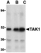
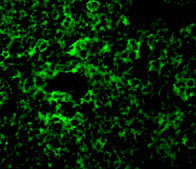
| Application Notes | TAK1 antibody can be used for the detection of TAK1 by Western blot at 1 to 4 µg/mL. For immunofluorescence start at 20 µg/mL. |
| Gene ID | 6885 |
|---|---|
| Other Names | TAK1 Antibody: TAK1, MEKK7, TGF1a, TAK1, Mitogen-activated protein kinase kinase kinase 7, Transforming growth factor-beta-activated kinase 1, TGF-beta-activated kinase 1, mitogen-activated protein kinase kinase kinase 7 |
| Target/Specificity | MAP3K7; |
| Reconstitution & Storage | TAK1 antibody can be stored at 4℃ for three months and -20℃, stable for up to one year. As with all antibodies care should be taken to avoid repeated freeze thaw cycles. Antibodies should not be exposed to prolonged high temperatures. |
| Precautions | TAK1 Antibody is for research use only and not for use in diagnostic or therapeutic procedures. |
| Name | MAP3K7 {ECO:0000303|PubMed:28397838, ECO:0000312|HGNC:HGNC:6859} |
|---|---|
| Function | Serine/threonine kinase which acts as an essential component of the MAP kinase signal transduction pathway (PubMed:10094049, PubMed:11460167, PubMed:12589052, PubMed:16845370, PubMed:16893890, PubMed:21512573, PubMed:8663074, PubMed:9079627). Plays an important role in the cascades of cellular responses evoked by changes in the environment (PubMed:10094049, PubMed:11460167, PubMed:12589052, PubMed:16845370, PubMed:16893890, PubMed:21512573, PubMed:8663074, PubMed:9079627). Mediates signal transduction of TRAF6, various cytokines including interleukin-1 (IL-1), transforming growth factor- beta (TGFB), TGFB-related factors like BMP2 and BMP4, toll-like receptors (TLR), tumor necrosis factor receptor CD40 and B-cell receptor (BCR) (PubMed:9079627, PubMed:16893890). Once activated, acts as an upstream activator of the MKK/JNK signal transduction cascade and the p38 MAPK signal transduction cascade through the phosphorylation and activation of several MAP kinase kinases like MAP2K1/MEK1, MAP2K3/MKK3, MAP2K6/MKK6 and MAP2K7/MKK7 (PubMed:8663074, PubMed:11460167). These MAP2Ks in turn activate p38 MAPKs and c-jun N- terminal kinases (JNKs); both p38 MAPK and JNK pathways control the transcription factors activator protein-1 (AP-1) (PubMed:8663074, PubMed:11460167, PubMed:12589052). Independently of MAP2Ks and p38 MAPKs, acts as a key activator of NF-kappa-B by promoting activation of the I-kappa-B-kinase (IKK) core complex (PubMed:8663074, PubMed:12589052). Mechanistically, recruited to polyubiquitin chains of RIPK2 and IKBKG/NEMO via TAB2/MAP3K7IP2 and TAB3/MAP3K7IP3, and catalyzes phosphorylation and activation of IKBKB/IKKB component of the IKK complex, leading to NF-kappa-B activation (PubMed:10094049, PubMed:11460167). In osmotic stress signaling, plays a major role in the activation of MAPK8/JNK1, but not that of NF-kappa-B (PubMed:16893890). Promotes TRIM5 capsid-specific restriction activity (PubMed:21512573). Phosphorylates RIPK1 at 'Ser-321' which positively regulates RIPK1 interaction with RIPK3 to promote necroptosis but negatively regulates RIPK1 kinase activity and its interaction with FADD to mediate apoptosis (By similarity). Phosphorylates STING1 in response to cGAMP-activation, promoting association between STEEP1 and STING1 and STING1 translocation to COPII vesicles (PubMed:37832545). |
| Cellular Location | Cytoplasm. Cell membrane; Peripheral membrane protein; Cytoplasmic side. Note=Although the majority of MAP3K7/TAK1 is found in the cytosol, when complexed with TAB1/MAP3K7IP1 and TAB2/MAP3K7IP2, it is also localized at the cell membrane |
| Tissue Location | Isoform 1A is the most abundant in ovary, skeletal muscle, spleen and blood mononuclear cells. Isoform 1B is highly expressed in brain, kidney and small intestine. Isoform 1C is the major form in prostate. Isoform 1D is the less abundant form |

Thousands of laboratories across the world have published research that depended on the performance of antibodies from Abcepta to advance their research. Check out links to articles that cite our products in major peer-reviewed journals, organized by research category.
info@abcepta.com, and receive a free "I Love Antibodies" mug.

Provided below are standard protocols that you may find useful for product applications.
Background
TAK1 Antibody: TAK1 (TGF-beta activated protein kinase 1) is a widely expressed enzyme originally identified as a mitogen-activated protein kinase kinase kinase (MAP3K7). It has since been shown to mediate various intracellular signaling pathways, such as those induced by TGF-beta and members of the Toll-IL-1R (TIR) superfamily, thus acting as an intermediate in both proliferative and innate and adaptive immune responses. TAK1 is normally present in cells in a complex with TAK1-binding protein 1 (TAB1) and either TAB2 or TAB3. Association with TAB1 triggers TAK1 kinase activity by inducing TAK1 autophosphorylation, while TAB2 and possibly TAB3 may contribute to SAPK2a/p38a-mediated feedback inhibition of TAK1 activity.
References
Yamaguchi K, Shirakabe K, Shibuya H, et al. Identification of a member of the MAPKKK family as a potential mediator of TGF-β signal transduction. Science 1995; 270:2008-11.
Irie T, Muta T, and Takeshige K. TAK1 mediates an activation signal from toll-like receptor(s) to nuclear factor-κB in lipopolysaccharide-stimulated macrophages. FEBS Lett. 2000; 467:160-4.
Akira S and Takeda K. Toll-like receptor Signalling. Nat. Rev. Immunol. 2004; 4:499-511.
Jiang Z, Ninomiya-Tsuji J, Qian Y, et al. Interleukin-1 (IL-1) receptor-associated kinase-dependent IL-1-induced signaling complexes phosphorylate TAK1 and TAB2 at the plasma membrane and activate TAK1 in the cytosol. Mol. Cell Biol. 2002; 22:7158-67.
If you have used an Abcepta product and would like to share how it has performed, please click on the "Submit Review" button and provide the requested information. Our staff will examine and post your review and contact you if needed.
If you have any additional inquiries please email technical services at tech@abcepta.com.
Ordering Information
Other Products
Shipping Information
