


 Foundational characteristics of cancer include proliferation, angiogenesis, migration, evasion of apoptosis, and cellular immortality. Find key markers for these cellular processes and antibodies to detect them.
 The SUMOplot™ Analysis Program predicts and scores sumoylation sites in your protein. SUMOylation is a post-translational modification involved in various cellular processes, such as nuclear-cytosolic transport, transcriptional regulation, apoptosis, protein stability, response to stress, and progression through the cell cycle.
 The Autophagy Receptor Motif Plotter predicts and scores autophagy receptor binding sites in your protein. Identifying proteins connected to this pathway is critical to understanding the role of autophagy in physiological as well as pathological processes such as development, differentiation, neurodegenerative diseases, stress, infection, and cancer.
SHANK3 Antibody
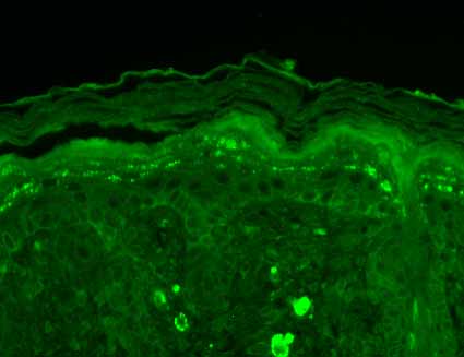
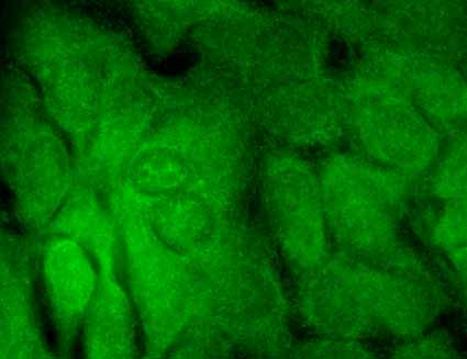
SHANK3 Antibody, Clone S69-46
- SPECIFICATION
- CITATIONS
- PROTOCOLS
- BACKGROUND

Application
| WB, IHC, ICC, IP, AM |
|---|---|
| Primary Accession | Q9JLU4 |
| Other Accession | NP_067708.1 |
| Host | Mouse |
| Isotype | IgG2b |
| Reactivity | Human, Mouse, Rat |
| Clonality | Monoclonal |
| Description | Mouse Anti-Rat SHANK3 Monoclonal IgG2b |
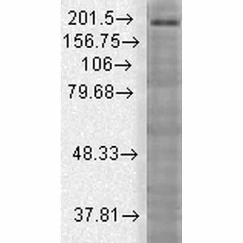
| Target/Specificity | Detects ~190kDa. No cross-reactivity against Shank1 or Shank2. |
| Other Names | AI841104 antibody, DEL22q13.3 antibody, KIAA1650 antibody, Proline rich synapse associated protein 2 antibody, Proline-rich synapse-associated protein 2 antibody, ProSAP2 antibody, PSAP2 antibody, SH3 and multiple ankyrin repeat domains 3 antibody, SH3 and multiple ankyrin repeat domains protein 3 antibody SH3/ankyrin domain gene 3 antibody, SHAN3_HUMAN antibody, Shank postsynaptic density protein antibody, Shank3 antibody, Shank3b antibody, SPANK 2 antibody, SPANK2 antibody |
| Clone Names | S69-46 |
| Immunogen | Synthetic peptide amino acids 840-857 of rat Shank3 |
| Purification | Protein G Purified |
| Storage | -20ºC |
| Storage Buffer | PBS pH7.4, 50% glycerol, 0.09% sodium azide |
| Shipping Temperature | Blue Ice or 4ºC |
| Certificate of Analysis | 1 µg/ml of SMC-336 was sufficient for detection of Shank3 in 10 µg COS cell lysate transiently transfected with Shank3 by colorimetric immunoblot analysis using goat anti-mouse IgG:HRP as the secondary antibody. |
| Cellular Localization | Cytoplasm | Cell Junction | Synapse | Postsynaptic Cell Membrane | Postsynaptic Density |

Thousands of laboratories across the world have published research that depended on the performance of antibodies from Abcepta to advance their research. Check out links to articles that cite our products in major peer-reviewed journals, organized by research category.
info@abcepta.com, and receive a free "I Love Antibodies" mug.

Provided below are standard protocols that you may find useful for product applications.
Background
Shank proteins make up a family of scaffold proteins identified through their interaction with a variety of membrane and cytoplasmic proteins (1). Shank proteins at postsynaptic sites of excitatory synapses play roles in signal transmission into the postsynaptic neuron. Shank proteins are also crucial in receptor tyrosine kinase signaling; specifically, Shank3 can mediate Erk-MAPK and P13K signaling which is crucial for tubule formation (2). Shank3 is also one of the latest genes to be associated with autism. A mutation of a single copy of Shank3 on chromosome 22q13 can result in language and/or social communication disorders (3).
References
1. Sheng M., and Kim E. (2000) Journal of Cell Science. 113: 1851-1856.
2. Schuetz G., et al. (2004) JCB. 167(5): 645-952.
3. Durand C.M., et al. (2007) Nature Genetics. 39: 25-27.
If you have used an Abcepta product and would like to share how it has performed, please click on the "Submit Review" button and provide the requested information. Our staff will examine and post your review and contact you if needed.
If you have any additional inquiries please email technical services at tech@abcepta.com.
Ordering Information
Other Products
Shipping Information
